
認知健康風險篩查
視力中心繼視網膜圖像分析系統(Automatic Retinal Image Analysis System,ARIA-Stroke Risk,ARIA腦中風風險指數檢測) 後,又再次得到香港中文大學授權使用其新開發的(ARIA-eWMH Cognitive Health Risk,ARIA認知健康風險篩查)平台,合作提供認知健康風險篩查服務。

認知障礙症 (Dementia)
於早年被稱為老人癡呆症的認知障礙症是因大腦神經細胞病變而引致大腦功能衰退的疾病, 病因可分為三大類:
-
-
-
-
-
阿兹海默症 - 成因未明,可能與遺傳有關。
-
血管性認知障礙症 - 因多次輕微中風令腦部受損而成。
-
其他會令患者有認知障礙症病徵的情況 - 腦積水、腦部感染、甲狀腺分泌不足、藥物中毒及抑鬱症等。
-
-
-
-
由於患者大腦神經細胞病變,而引致大腦功能衰退,患者的記憶、理解、語言、學習、計算和判斷能力都會受影響,部分且會有情緒、行為及感覺等方面的變化、一般最先的影響是記憶,其實,我們間中都會忘記一些生活細節,例如特然忘記了某個字的冩法或串法,或是『昨晚食乜餸 』?的挑戰短期記憶的玩意!這和忘記老伴的名字,或忘記了家在何處的情況是有很大的分別。
 雖然現時還没有治愈認知障礙症的方法,但主動管理可改變危險因素、可以推遲或延缓認知障礙症的病發或進展。因為治療及照顧腦退化及患者,不單是患者需要配合照顧,其家人都需要面對經濟、情緒及生活上的沉重壓力。如能及早日發現並配合適當冶療,能減緩退化,降低病徵對家庭的影響,維持一定的生活質素。
衞生署長者健康服務聯同中文大學醫學院精神科學系於2005/06的研究顯示,在本港70歲以上的長者中,約有9.3%患有認知障礙症,男性的患病比率為8.9%,女性則為15.3%。
認知障礙症亦是一個迅速擴大的全球公共衛生問題。
跟據世界衛生組織的統計,隨着人口老化,認知障礙症患者愈來愈多,全球大約有 5 千萬人患癡呆症,其中 60%生活在低收入和中等收入國家或地區,全球每年新增的病例接近一千萬,需要被照顧的人仕與日俱增,社會各方面壓力亦驟增,據世衛估計,預計到 2030 年全球痴呆症患者總數將達到 8200 萬,2050 年將達到 1.52 億例。癡呆症導致政府、社區、家庭和個人費用增加,各經濟體生產率下降。在2015 年,癡呆症的全球社會總成本估計為8180 億美元,相當於全球國內生產總值的 1.1%。估計到2030年,用於照顧認知障礙症患者的費用,將會達至每年弍萬億美元,這當然會為各個經濟體的財政帶來影響。
ARIA認知健康風險篩查
認知障礙症亦是老年人致殘和依賴的一個主要原因,可能會破壞病人、其護理者和家庭的生活質素。
若能及早篩查出高風險認知障礙症患者,並作出適當治療或預防措施,從而減輕各方面的負擔,就能夠改善認知障礙症患者,及其照顧者的生活質素。
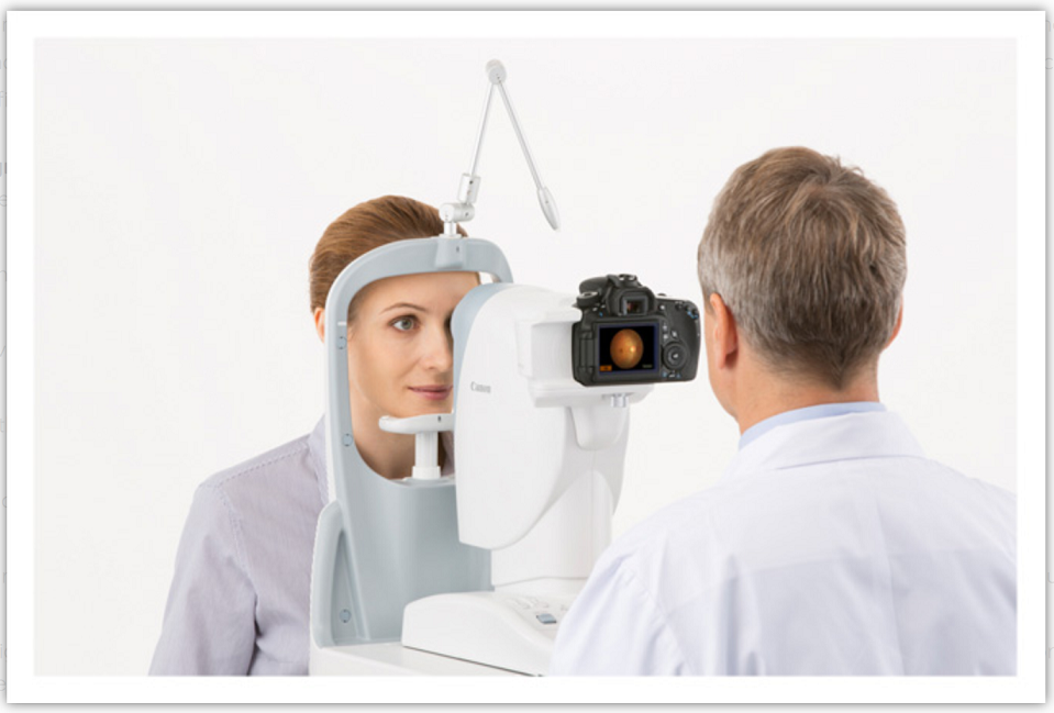
必需要驗眼
ARIA-eWMH 的基本運作,與視網膜圖像的擷取影像的要求跟ARIA-Stroke Risk 一樣,為了要保持視網膜圖像的的可靠性,視光師會為應診者先作全面的眼睛健康檢查,證實眼睛健康正常,然後在瞳孔放大的情況下進行視網膜圖像擷取,這樣的檢查及擷取的影像質素才有保證,用作分析及評估應診者患上認知障礙症風險的高低,準確度可高達90%以上。
甚麼人仕適合使用ARIA-eWMH ARIA認知健康風險篩查
-
-
-
-
-
-
50歲或以上
-
沒有中風及\或輕微中風的病歷
-
沒有被診斷為認知障礙或輕微認知障礙
-
視網膜圖像清晰
-
-
-
-
-
ARIA視網膜圖像分析系統操作相對簡單,唯一要求是要拍攝到清晰及一致性的的視網膜影像。檢查屬非入侵性,連續性檢查亦不會影響眼睛,亦可作隨機性檢查,不受時間或地域限制;不帶輻射,無需抽血,檢查結果亦不會因年齡,表達能力,教育程度或文化差異而受應響!費用相對便宜。
ARIA視網膜圖像分析系統的作用是風險評估,無法診斷病症,用戶若知悉自己是高危人士後,可做進一步的檢查,或改變其不良生活習慣,從而對症下藥,建立健康的生活模式,減低疾病對生活質素的影響。
查詢或預約
油麻地診所 : (852) 2385-6068
.
旺 角 診 所 : (852) 2396-6629
.
有關 Automatic Retinal Image Analysis for estimated White Matter Hyperintensities (ARIA-eWMH)
ARIA-eWMH是利用全自動視網膜圖像分析方法 (ARIA) 並以腦磁共振成像(MRI)作為分析標準,計算腦內是否有嚴重的年齡性白質病變(ARWMH)風險,這技術有超過90%的靈敏度和特異性。[1] 腦內白質病變(WMH)是腦小血管疾病的結果。WMH量與多個認知領域顯著相關,包括記憶,概念化和視覺實踐技能 [2, 3] 。縱向研究證實,白質病變的惡化導致腦萎縮和認知能力下降。[4] 從一些重複MRI研究所示,WMH可能隨著時間增加也有可能減少 [5, 6] 。WMH被證明是阿爾茨海默病的核心特徵,WMH體積可能在預期症狀發作前約5-6年開始出現。腦β-澱粉樣蛋白被認為是阿爾茨海默病的合理解釋之一,與臨床前階段的WMH體積(p = 0.03)和載脂蛋白E(APOE)ε4狀態(p = 0.002)顯著相關[10]。
ARIA-eWMH was developed to assess the risk of severe age-related white matter hyperintensities (ARWMH) in the brain based on automatic retinal images analysis (ARIA), using cerebral magnetic resonance imaging (MRI) as gold standard and achieved more than 90% sensitivity and specificity. [1] White matter hyperintensities (WMH) in the brain are the consequence of cerebral small vessel disease (SVD). WMH volume significantly associated with multiple cognitive domains, including memory, conceptualization, motor abnormalities and visual practical skills. [2, 3] Longitudinal studies have confirmed that worsening of white matter lesions contributes to brain atrophy and cognitive decline. [4] WMH may progress but it may also regress along time as shown by repeated MRI studies. [5, 6] WHM was shown to be a core feature of Alzheimer’s disease and WMH volumes may start to appear approximately 5-6 years prior to expected symptom onset. [7, 8, 9] Cerebral beta-amyloid protein, which is believed to be one of the plausible explanation of Alzheimer’s disease, is significantly associated with WMH volume (p=0.03) and apolipoprotein E (APOE) ε4 status (p=0.002) in the preclinical stage.[10]
免責聲明:
Automatic Retinal Image Analysis for estimated White Matter Hyperintensities (ARIA-eWMH)
認知健康風險篩查: 視網膜圖像分析系統分析報告主要目的是為了促進健康,並不能替代專業醫療諮詢。凡接受任何治療之前,或對治療有任何問題,請諮詢專業的醫護人員。
Note: Automatic Retinal Image Analysis for estimated White Matter Hyperintensities (ARIA-eWMH)
Cognitive Health Risk level is not intended for disease diagnosis or therapeutic purposes. It is not intended to replace regular eye examinations or medical examination. The information on Stroke Risk Level is provided to the clients for health promotion ONLY and use as a reference. It is neither intended nor implied to be a substitute for professional medical advice. Always seek the advice of medical professionals or other qualified healthcare providers prior to starting any new treatment or address questions your clients may have regarding a medical condition.
參考資料
Risk reduction of cognitive decline and dementia - WHO Guidelines
ARIA腦中風風險指數檢測
References
-
Lau A, Mok V, Lee J, Fan Y, Zeng J, Lam b, Wong A, Kwok C, Lai M, Zee B, “Retinal image analytics detects white matter hyperintensities in healthy adults”, Annals of Clinical and Translational Neurology, Volume 6, Issue 1, January 2019, Pages 98-105
-
Inzitari D, Pracucci G, Poggesi A, et al. “Changes in white matter as determinant of global functional decline in older independent outpatients: Three years follow-up of LADIS (leukoaraiosis and disability) study cohort”. BMJ Clinical research 2009; 339: b2477
-
Prins ND, Scheltens P, “White matter hyperintensities, cognitive impairment and dementia: an update”, Nat Rev Neurol. 2015 Mar;11(3):157-65
-
Schmidt R, Ropele S, Enzinger C, Petrovic K, Smith S, Schmidt H, Matthews PM, Fazekas F: “White matter lesion progression, brain atrophy, and cognitive decline: The Austrian stroke prevention study”. Ann Neurol 2005;58:610-616
-
Cho AH, Kim HR, Kim W, Yang DW “White Matter Hyperintensity in Ischemic Stroke Patients: It May Regress Over Time”, Journal of Stroke 2015; 17(1): 60-66.
-
Ramirez J, McNeely AA, Berezuk C, Gao F, and Black SE, “Dynamic Progression of White Matter Hyperintensities in Alzheimer’s Disease and Normal Aging: Results from the Sunnybrook Dementia Study”, Frontiers in Aging Neuroscience, March 2016, Volume 8, Article 62.
-
Brickman AM “Contemplating Alzheimer’s disease and the contribution of white matter hyperintensities”, Curr Neurol Neurosci Rep (2013) 13:415
-
Lee S, Viqar F, Zimmerman ME, Narkhede A, et al.; Dominantly Inherited Alzheimer Network, “White matter hyperintensities are a core feature of Alzheimer’s disease: Evidence from the dominantly inherited Alzheimer network”, Ann Neurol. 2016 Jun;79(6):929-39.
-
Araque Caballero MÁ, Suárez-Calvet M, Duering Met al., “White matter diffusion alterations precede symptom onset in autosomal dominant Alzheimer’s disease”, Brain 2018 Oct 1;141(10):3065-3080
-
Kandel BM, Avants BB, Gee JC, McMillan CT, Erus G, Doshi J, Davatzikos C, Wolk DA, “White matter hyperintensities are more highly associated with preclinical Alzheimer’s disease than imaging and cognitive markers of neurodegeneration”, Alzheimers Dement (Amst). 2016 Apr 7;4:18-27